Excitable Cells and Biomechanics
Research Areas
We have a shared interest in excitable cells and contractile tissue. The research in this group falls into two main categories: Excitable Cells and Biomechanics.
Exitable Cells
The molecular basis for the transport of ions, water and nutrients, ion channels, electrophysiology, pharmacological treatment, action potentials, cardiac arrhythmias, epilepsy, migraine, evolution of the electrical signaling in the mammalian heart, stem cells derived organoids
Biomechanics
Fascia, locomotor system, biomechanics, lateral raphe, thoracolumbar fascia, acoustic myography, multi-frequency bioimpedance, isolated tissue bath studies, rheumatology, sports medicine, geriatrics, structural anatomy and pathology.
Our missions are
- To understand the structure, function and regulation of ion channels, receptors and transporters and identify novel pharmaceutical treatments
- Understand the conductive system and the role of ion channels in excitation-contraction coupling in healthy and diseased hearts
- Advance the understanding of the skeletal muscle, fascia and locomotor system to improve diagnostics, treatment and rehabilitation in companion animals
- To measure muscle function and health in both static and dynamic situations
- To deliver state-of-the-art research within these fields and to incorporate it in our teaching
- We have an established electrophysiological profile and the labs has a wide variety of equipment to measure function of transport molecules at single molecule (purified ion channel in lipid bilayers), whole cell level (two-electrode voltage-clamp and patch-clamp), organoid and organ level (membrane potential measurements, ECG, Ussing chamber, bio-impedance). We will strengthen this profile and use our expert skills in the fields to increase collaboration with university partners and industry.
- By increasing the understanding of the conductive system and electrical signaling in the mammalian hearts, we aim to refine the use of animal models. Pigs and dogs are extensively used as models for humans in drug development and cardiac safety pharmacology, however, to properly design animal studies and interpret the results, an in-depth knowledge about their anatomy and electrophysiology is crucial and presently insufficient.
- Establish organoids based on animal and human inducible pluripotent stem cells (iPSC) to evaluate genetic variants and the effects of pharmaceutical treatment. We have established collaborations with clinicians in human and animal practice. Together with Kristine Freude’s group, we aim to provide excellent stem cell based in vitro models of primarily cardiac, but potentially also other tissues, that can be used to investigate pathological changes associated with different diseases as well as to test pharmaceutical treatment (personalized medicine). Our in vitro models can be validated by in vivo models in EAM and our aim is to create cell based organoids that replicate human and animal tissue to be used in drug development and safety pharmacology and thereby reduce the number of experimental animals.
- There is an unmet need for diagnostics and treatment of injuries in the locomotion system. Our vision is to establish an International Center of Excellence for Biomechanics, Training and Rehabilitation for horses and other animals at the Department of Veterinary and Animal Sciences
- To develop novel techniques that can be applied in both the Veterinary and Human clinics
- To establish a synergy between clinical genetics, organoid models and in vivo research to facilitate personalized medicine
- Improve our research through collaboration with partners in academia, clinics and industry
Excitable Cell Research
We are specialized in electrophysiological characterization of ion channels, transporters and receptors. Ion channels, transporters and receptors are important for cell homeostasis, intracellular signaling as well as maintenance of the membrane potential and action potential generation. Malfunction of transporters and ion channels e.g. due to genetic variants, can result in diseases, such as migraine and cardiac arrhythmias. Their important function in cellular function makes them potential drug targets for treatment of e.g. cardiac disease, migraine and parasitic infections.
Current Projects in Molecular Physiology (Professor Dan Klærke)
 Crystal structure and function of ion channels and transporters. We record currents from single ion channels expressed in artificial lipid bilayers to test the functionality of ion channel proteins purified from Saccharomyces cerevisiae
- Identification and characterization of novel ion channel or receptors in malaria and other parasites and test of pharmaceutical treatment
- Elucidation of the mechanism linking cell swelling to activation of Slick and Slack channels
Professor Dan Klærke, e-mail: dk@sund.ku.dk
Current projects in Cellular Electrophysiology (Kirstine Callø)
- We have a special interest in cardiac electrical signaling in normal and diseased hearts. In collaboration with cardiologists we have identified genetic mutations in ion channel genes in human and animal patients with cardiac arrhythmia. We perform electrophysiological characterizations of these mutated ion channels to improve diagnostic work and treatment. Recently we have established cardiac organoids based on induced pluripotent stem cells (iPSC).
- Development and evolution of the conductive system and electrical signaling in mammalian hearts. In this project we map the conduction system of hearts and obtain ECGs from different mammalian species to improve the understanding of the excitation-contraction process in large hearts.
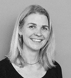 Associate professor Kirstine Callø (Cand. Scient. Human biology, PhD, Dr Vet Sc.). Kirstine is an expert in ion channel function and electrical signaling in healthy and diseased hearts. Her research is focused on improving diagnostics and pharmaceutical treatment of human and animal patients. She is in particular interested in patients with cardiac arrhythmia and sudden cardiac death due to variants in genes encoding ion channels. To get a better understanding of the electrical signaling and cardiac contraction, Kirstine is investigating the cardiac conduction system and electrical activation in very large mammals.
E-mail: KirstineC@sund.ku.dk
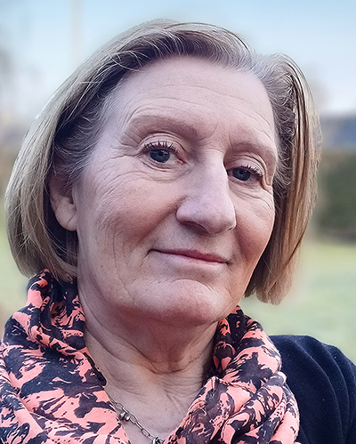 Vibeke Grøsfjeld Christensen (Pharmaconomist (Laboratory technician)):
Vibeke has extensive experience with laboratory work and has been involved in many different research projects. She is involved in the planning and execution of the experimental work and in processing the resulting data. Her experience in the laboratory includes ELISA, immunohistochemistry, qPCR, western blot, pressure myograph, molecular biological techniques, electrophysiology (TEVC, Orbit, Port a Patch) and cell culturing. She enjoys to supervise the students in the laboratory and to teach them good laboratory practice.
E-mail: vgc@sund.ku.dk
Biomechanics Research:
Up until quite recently biomechanical studies of the locomotion system in both the human and veterinary fields has been solely focused on muscle and bone connections thereby presenting joint flexibility and tendon health as the major structures in movement.
However, during the last decade biomechanical research in humans has focused more on connective tissue in its different forms, revealing these structures to be a forceful and essential contributor to the function of the locomotion system. It has also become apparent that these structures work in the body in a highly integrated, correlated, and complex way. Such discoveries as these serve to push and drive research into the locomotion system, inspiring us to investigate and more fully understand previously identified structures in a new light.
In studies performed by our research group (both veterinary and human fields) it is clear that very specialized and highly tuned myofascial interactions occur both at the macro- and microscopic level.
We are currently interested in a number of research projects, but some examples of our recent work include the thoracolumbar fascia (TLF), as well as the function and integration of this in relation to the biomechanics of the back.
In humans, an anatomical structure termed a lateral raphe has been identified, this is a place where several fascia layers meet. This lateral raphe has recently been identified as being a structure of great importance with regard to addressing the question of how forces are distributed over regions of the body. Of perhaps even greater importance is the finding that a dysfunctional raphe has been related to lower back pain.
In addition to the projects investigating connective tissue, we are also very interested in some basic research questions regarding muscle tissue function, anatomical structures and pathology. Measurements of muscle health are of great interest for various medical fields including neurology, rheumatology, sports medicine and geriatrics. We are particularly interested in the development of new non-invasive techniques such as acoustic myography and multi-frequency bioimpedance as well as the myoton.
Non-invasive assessments of muscle tissue in vivo allows the evaluation of therapeutic effects after an intervention without crossing the barrier of the skin and thus without disturbing the natural environment of the muscle. Currently we have several projects where we investigate muscle fatigue, muscle recovery and different muscle pathologies using mostly non-invasive techniques both in canine, equine and human models.
About us:
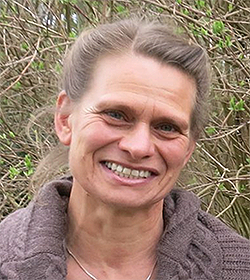 Dr Vibeke Sødring Elbrønd: (Veterinarian, PhD): Lecturer in Anatomy at the Faculty of Health & Medical Sciences, Copenhagen University. Vibeke has considerable expertise in the fields of fascia and associated structures, functional anatomy, the preparation of tissue, microscopy and the interpretation of images. Vibeke has a post doctoral education as a veterinary chiropractor, and is trained in connective tissue treatment, acupuncture as well as functionally based indirect techniques.
E-mail: vse@sund.ku.dk
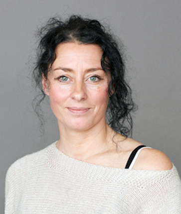 Dr Jessica Pingel: (Sports & Health, PhD): Associate Professor at the Department of Neuroscience, Copenhagen University. Jessica has worked with tendon-morphology, biochemistry, microvascular function, sports medicine and health, as well as more recently muscle pathophysiology after CNS lesions. Her expertise includes ELISA, qPCR, western blot, immunohistochemistry, transmission electron microscopy, second harmonic generation microscopy, sEMG, Dexa, MRI, contrast enhanced ultrasound, muscle force measurements, next generation sequencing, DNA profiling, SNP analysis and capillary electrophoresis. In addition to this Jessica speaks four languages.
E-mail: jpingel@sund.ku.dk
 Dr Adrian Paul Harrison: (Physiology, D.Phil): Lecturer in Animal Production Physiology at the Faculty of Health & Medical Sciences, Copenhagen University. Adrian is trained in agriculture, biochemistry & nutrition as well as possessing a D.Phil. in physiology from Cambridge University. His main research interest has been muscle, first at the level of porcine muscle development, later with isolated rat muscle contraction, and clinical studies of muscle function and weakness. Adrian´s research also covers intestinal function, GI tract motility, bone density and palaeo-biomedicine. Adrian is the inventor of acoustic myography.
E-mail: adh@sund.ku.dk
